


Camellia Seed Meal Provides the Starting Material
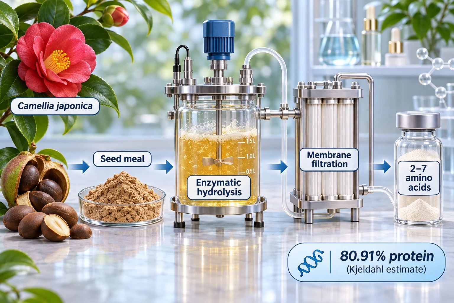
The study, identified as IFSCC 2026 paper 108, was conducted by Shanghai Forest Cabin Cosmetics Group and a joint anti-ageing laboratory at Shanghai Jiao Tong University, according to BeautyNews. The researchers used the solid meal remaining after oil extraction from Camellia japonica seeds. They extracted the seed proteins, broke them down with enzymes and used membrane filtration to obtain a fraction containing short peptides, predominantly two to seven amino acids long. The resulting preparation had a reported protein content of 80.91%, as estimated by the Kjeldahl nitrogen method. The process gives an oil-production by-product a potential use as a cosmetic ingredient feedstock.
Camellia Seed Meal Provides the Starting Material
The study, identified as IFSCC 2026 paper 108, was conducted by Shanghai Forest Cabin Cosmetics Group and a joint anti-ageing laboratory at Shanghai Jiao Tong University, according to BeautyNews. The researchers used the solid meal remaining after oil extraction from Camellia japonica seeds. They extracted the seed proteins, broke them down with enzymes and used membrane filtration to obtain a fraction containing short peptides, predominantly two to seven amino acids long. The resulting preparation had a reported protein content of 80.91%, as estimated by the Kjeldahl nitrogen method. The process gives an oil-production by-product a potential use as a cosmetic ingredient feedstock.

Matrix and Barrier Protein Signals Also Changed
The study used immunofluorescence to examine proteins associated with skin structure and epidermal differentiation. BeautyNews reported stronger signals for type I and III collagen and elastin alongside a weaker signal for matrix metalloproteinase-1 (MMP-1), an enzyme that can break down fibrillar collagen. Signals for type IV and VII collagen also increased, as did those for filaggrin and loricrin, two proteins associated with epidermal differentiation and barrier formation. The public summary described these changes qualitatively. The findings provide cellular readouts relevant to matrix and barrier research, while functional changes in intact skin remain to be tested.
Autophagic Flux Remains a Question for Follow-Up Studies
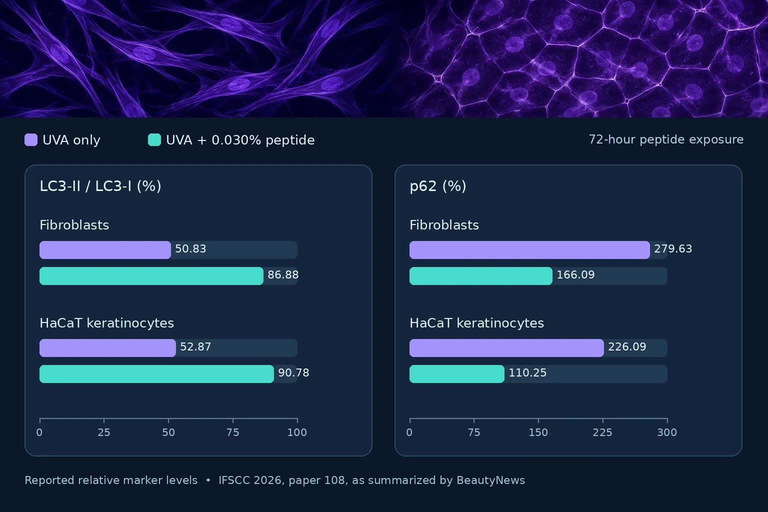
Autophagic flux describes how cellular material moves through the autophagy pathway and is ultimately degraded in lysosomes. The paired LC3 and p62 changes are consistent with improved turnover, but protein abundance at a single time point cannot establish how quickly material is being cleared. Comparing LC3-II turnover with and without lysosomal degradation blocked would help test that interpretation directly. BeautyNews also reported that the conference paper did not specify the UVA dose, an important detail for reproducing the experiment. The publicly reported findings come from cell cultures, with effects in human skin still to be evaluated.


